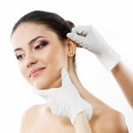
Otoplasty - protruding ears correction
Otoplasty
Price from 743 GBP
Otoplasty protruding ears correction is a purely aesthetic procedure. For the majority of cases, erectile ears are a psychological problem, especially among children, but the problem also affects adults. A large degree of ear deformity can lead to lower quality of life, reduced self-esteem, social exclusion and poor school performance
Otoplasty - protruding ears correction - the correction of protruding ears is a purely aesthetic procedure. For the majority of cases, erectile ears are a psychological problem, especially among children, but the problem also affects adults. A large degree of ear deformity can lead to lower quality of life, reduced self-esteem, social exclusion and poor school performance.
Children due to the problem of protruding ears are sometimes rejected by peers, ridiculed or even intimidated. In the case of adults, protruding ears for many people are the cause of complexes and ill-being. It often leads to social isolation, problems at work and closure. Adults may also have practical problems, for example with the assumption of specialized headgear for example a motorcycle helmet.
Correction of protruding ears
The problem of protruding ears affects about 1-5% of the population, in some cases it occurs in the family. Deformation can affect one or both pinna. More often, however, it occurs on both sides.
Outstanding ear surgery is one of the most common aesthetic procedures in the head area and the most common aesthetic procedure performed in children. There is also a 4-fold increase in the frequency of correction of the protruding auricles in the female sex. Dieffenbach made the first described correction of ear out in 1845.
The human ear is the organ of hearing and balance. It is composed of three components. Two of them are the middle and inner ear, which are located inside the skeletal system of the skull. The outer part is the ear mucus and the external auditory canal separated from the middle ear by the eardrum. The ear mantle is made of elastic cartilage covered with skin, which is unstoppable in relation to the ground. The shape of the ear primarily determines the cartilage skeleton. The frontal surface of the ear has no subcutaneous fat and is therefore particularly sensitive to injuries.
Construction of the pinna.
Despite the small size, the earlobe is made of many elements. The individual components are: a hoop, a branch of a hoop, a hollow tail, a dam, a leg, dams, a frontal gangrene, a triangular, a shuttlecock, a shell cavity, a shell boat, an earlobe, a piece, a slit, a subcutaneous incision, a front incision.
The outermost part is the cap that goes down into the earlobe (the only part is uncommunicative) downwards. To the front of the hollows there is a hue called a glaze. The gullet is separated from the hollow by the lenght, to the gland separates into two branches between which there is a triangular pit. The closest to the auditory canal are shells. A section is located forward of the auditory canal, which in the back and below is adjacent to the scraper separated by a subcutaneous incision. The most important elements are:
- Hem.
- Shuttle.
- Seashell.
- Earlobe.
- Scrap.
- ear
The ear piece in an adult differs from the children mainly due to its plasticity. Children's ears are softer and more plastic than adult ears. Elder cartilage is more calcified and rigid, which is why it is more difficult to correct the outlier ears in adults. After 5 years of age, the ear can grow very slowly, which is why this period, according to some surgeons, is the best for any correction of the ear, including the correction of the ears outliers. Some operators believe that the best period to carry out this surgery is between 10 and 14 years of age, as the earlobe ceases to grow completely during this period.
All the components of the pinna are on the front surface of the ear. The back surface of the pinna is the "negative" of the front surface. During the correction of the outlier ears, the shape of the front surface is changed by modeling the cartilage, but the cuts are made on the back surface to hide the scars.
What are the features of normal ear pinna?
The most important features of the auricle are its size, location and shape.
The size of the outer ear refers to the corresponding elements on the face. The correct size is the upper edge of the pinna, located at the height of the eyebrows and the lower pole of the pinna, located at the height of the passage of the nose column into the upper lip.
The position of the ear is assessed in several planes, lateral, upper and upper. Looking from the side, the correct position of the pinna is determined by assessing the position of the long axis of the ear in relation to the frontal plane and the angle between these planes should be around 30 '. When viewed from the front, the suture should properly extend to a maximum of 1.5 cm from the lateral plane of the head. In turn, looking from the top angle between the earlobe and the surface of the head is about 30 ', the angle between the shell and the shuttle is 90'.
When making the correction of the outlier ears, one should adhere to the anatomical position of the ear in order not to make an excessive correction, which could make the ears look flat.
The benefits of correcting outward ears
Outstanding ear surgery is a procedure in the field of aesthetic surgery. The main assumption and benefits resulting from the operation is to improve the patient's mental comfort. Some people have practical problems associated with protruding ears, for example when setting up a specialized headgear (helmet, helmet). In such situations correction of outliers can also bring benefits in the professional sphere. The operation is relatively short and you do not have to stay in the hospital after it. The treatment gives satisfactory and long-lasting effects.
Indications for correction of protruding ears
A surgeon or otolaryngologist performs the qualification for surgery for the earlobes. The main indication for surgery is a significant deviation from the natural, anatomical shape and position.
The outlier ear correction should be performed when there is a significant deformation, because only then are the effects of the operation visible.
The treatment is indicated in people whose ear deformation leads to a decrease in the quality of life due to complexes affecting self-esteem, mental health and physical activity and social activity.
Contraindications to the correction of protruding ears
The main contraindications to the correction of protruding ears are:
- infectious diseases;
- general inflammation;
- locally occurrence of inflammation, purulent lesions;
- tendency to form keloids;
- anticoagulants (discontinue or change 7 days before)
- mental state of the patient - the surgeon may not undertake an operation when he / she thinks that the deformation of the pinches is not large enough. The cosmetic effect obtained after correction may be too small and unsatisfactory. Some patients have a greater psychological than aesthetic problem, despite the person's correction of the ears protruding in a technically perfect way, they may not be satisfied with the result.
Before surgery, correction of protruding ears
During consultations before the procedure of correcting the outlier ears, the doctor collects the interview and examines the patient. It is also the best time to ask questions to your doctor. It is important then to inform the doctor about all diseases, medicines and allergies you have. People taking anticoagulants must, according to the doctor's instructions, set them aside one week before the procedure or switch to another anticoagulant with a lower potency.
The surgeon may also order a medical consultation with another specialist, such as a cardiologist if the patient has a chronic heart disease. During the consultation, the type of anesthesia to which the patient will be subject is also determined. In the case of correction of the outlier ears, it is usually local anesthesia.
Before surgery, consent for the surgery is signed. When signing consent, the doctor informs the patient about how the procedure will be performed, what will be done, what are the possible complications and other treatments.
During the consultation before the operation of correcting the ears, the surgeon also assesses the degree and type of auricular deformation, their shape, size and location. Photographs are taken for documentation.
Type of anesthesia for the correction of protruding ears
Anesthesia used when correcting outliers is typically local anesthesia. A small amount (a few to a dozen or so milliliters) of a mixture of lignocaine and adrenaline is used for anesthesia. The needle is inserted into the anterior and posterior surface of the pinna, and the behind-the-ear region over the head to inject the area of nerves innervating the turbinate. Sometimes, during anesthesia of the ear, a temporary paralysis of the facial nerve may occur.
What does the correction treatment of the ears look like?
During the correction of the outlier ears, the shape of the front surface is changed by modeling the cartilage. The cut is made on the back surface to hide the scar. The hollows of the anterior surface of the pin correspond to the projections on the posterior surface and, in turn, the recesses in the back correspond to the hares in the front. During the correction of the outlier ears, it is most important to identify the elements of the posterior surface and the corresponding elements in the front, therefore during the operation piercing the ears with needles with ink.
There are over 60 types of ways to correct outliers. The following article discusses the most typical procedure. It should be remembered that the surgeon, during consultations in the pre- and intraoperative period, chooses the way he will operate. Qualification for a given type of surgery is based on the clinical situation, the nature of the deformity, the age of the patient and the experience of the operator.
Ear corrections are made by approximating the corresponding cartilage points on the front surface through the seams made on the back surface. In this way, a dam is formed and the turbinate withdraws. In addition, if the vein is too large, you can cut a part of the cartilage.
The soft cartilage can be formed using only sutures. When we deal with rigid cartilage, cuts are made at selected places to weaken and plasticize it.
During the correction of the ears, protruding cuts are made on the back surface of the ear, only the planning is made on the front surface. The skin is initially cut on the back of the pinna at a distance of about 1 cm from the posterior furrow. Then, if need arises, part of the cartilage is cut out. A convexity of the dam is then formed by appropriate bending of the cartilage, which is stitched with insoluble threads. In adults, you can also make cartilage cuts in the right places to make it plastic, in children the cartilage is soft and usually does not require a maneuver. Sometimes the earlobe is sewn to the fascia of the head, and the earlobe may also be reduced during the procedure. After proper cartilage placement, excess skin is cut and sutured with absorptive or insoluble threads.
After correcting the outlier ears, the appropriate dressing is performed. The treated area is covered with a fat dressing (with sterile vaseline) and properly formed gases, and the whole is supported with a bandage. The dressing is designed to press the treated pinna properly to prevent the formation of a hematoma. On the other hand, it protects the operated ear from injuries and excessive stress, for example during sleep. The earlobe after protruding ears looks the most physiologically when the labrum is immediately behind the dam without excessive deviation.
The duration of the treatment is 30-45 min.
Time and course of convalescence after correction of protruding ears
On the day of surgery, correcting ears outliers if local anesthesia was applied, the patient can go home. Before leaving, he should receive an information card with all recommendations. The most important is to follow the instructions of the attending physician, because he knows the clinical situation of the patient best.
In the first day a special dressing is put on, which should be changed every day. In the first days after correction of the ears, the emerging pain in the surgical site may be so strong that it will be necessary to use painkillers. If skin sutures have been applied, they should be removed 5-7 days after surgery. It is recommended to clean the head after removing the sutures, before downloading them, make sure that the wounds do not wet, but only rinse and wipe dry. It is recommended to wear a flexible dressing or a headband to protect your ears, especially during sleep.
The first inspections after correction of the ears outgoing for a period of about 2 weeks can take place even every few days. For the first 1-2 weeks after surgery, you should also avoid effort and lead a saving lifestyle.
The skin overgrows within a few days, however, it should be remembered that the scar in the human body is formed from six months to a year, so it should be remembered that at this time the strength of the operated site may be weaker.
The effects of correction of outliers
The correction of the outlier ears leads to an improvement in the symmetry, proportion and size of the earlobes. Changing the appearance of the ears has an impact on improving the appearance of the entire face and improving the mental comfort of the patient. The greater the deformation, the better the final result and the patient's satisfaction. When the deformation of the ears is very small, the effect of the surgery may be imperceptible to the patient, therefore the surgeon may not undertake such a procedure.
- Recommendations after correction of protruding ears.
- Recommendations after the correction of protruding ears are:
- follow the instructions of the attending physician;
- putting on checks at set times;
- daily dressing change;
- prohibition of soaking wounds - worse wound healing, higher probability of infection;
- wound cleaning with skin disinfectant - skinsept, octenisept;
- use of oral analgesics in case of intensification of symptoms;
- use of a elastic band to protect the ear;
- avoiding physical activity before 1-2 weeks after surgery;
- photo of stitches on the 5-7 day after surgery;
- avoiding contact sports for the next 2-3 months
- in the event of deterioration of the local condition, eg excessive swelling, redness, appearance of purulent secretion or the appearance of other disturbing symptoms, try to get to the doctor who performed the procedure. If this is not possible, report to the nearest surgical IP.
How long do the effects of correction of the outward ears persist?
If in the early period there were no complications and recurrence, the effect of correcting ears outliers may persist for the rest of their lives.
How to avoid complications after correcting ears outliers
- follow the doctor's instructions,
- put on set inspection dates,
- take care of the hygiene of the surgical site, change the dressing daily, wash the wound with octenisept, scinsept or soapy water,
- it is forbidden to soak the operated place, wash preferably in the shower and immediately before changing the dressing,
- use specialized dressings that properly tighten the ear and at the same time do not cause excessive pressure,
- do not bend the ear mites within a few weeks after the procedure, always protect the ears to sleep (dressing or bandage),
- use a tourniquet thanks to which the ear cone will properly adhere to the head,
- if any disturbing symptoms appear, such as a sudden appearance of fever, redness, excessive heat or oil in the surgical site, report immediately to the surgeon performing the procedure or the nearest Surgical Reception Chamber,
- avoid effort for about 2 weeks,
- for 2-3 months, avoid contact sports.
Possible complications
The operation is considered to be safe, statistically, in the course of ear correction, outliers complications are very rare. However, like any procedure, this one is also subject to certain risks of complications.
In the technique with the use of a thread, the cartilage can be perforated at the thread passage, i.e. the so-called fistula is formed. Another complication is the intersection of the cartilage or the unsightly position of the skin, the folds above the threads or the distortion of the pinna.
Sometimes minor cartilage deformations may be unnoticed during the operation of correcting the outlier ears and become visible only after the procedure. Reoperation may be required, which is best done within 1-2 weeks of the previous procedure. Other early and late complications include:
- pain - do not use any medicines containing aspirin and ibuporofen, it is best to use pyralgine or paracetamol
- bleeding, hematoma - a re-operation may be required to stop the bleeding or to remove the hematoma
- Sensations of the pinna tenderness - usually transient
- decubitus-prevented by appropriate formation of dressings
- inflammation, infection - general action may be required - antibiotic and local. These complications often cause secondary deformation of the turbinate
- narrowing of the auditory canal - temporarily as a result of edema, prolonged as a result of cartilage deformation, reoperation may be required
- asymmetry - reoperation may be required
- relapse - partial or complete - usually 1/3 of the upper ear
- scarring - means an overgrown, pathological scar of a characteristic red color, excessive remodeling, overhanging and giving an unpleasant sensory experience. The three most frequent locations of keloids are the chest, shoulders and ears, therefore, in the person with which he has had a cripple, there is a high probability that the change will appear after correcting the ears.
Recommended additional treatments
During the correction of the outlier ears, the other ear structures can be corrected at the same time, eg reduce the earlobe, enhance or reduce the dam, shuttle, etc. If necessary, the external auditory canal or the tympanic membrane can also be plasticized.
Price
from 743 GBP
Duration
30-45 min
Anesthesia
local anaesthesia
Hospitalisation
not required
Recovery time
about 2 weeks
Effects
after 2 weeks
Lenght of the effects of the treatment
permanent
Beauty Group
71-476 Szczecin
Otoplasty - protruding ears correction:
from 1273 GBP
Price list of the treatment
We encourage you to familiarize yourself with the prices of the procedure Otoplasty - protruding ears correction in the city chosen by you. The price depends on the scope, method of the procedure, type of anesthesia as well as the location and reputation of the clinic.
We encourage you to take advantage of free assistance in arranging a private visit to the clinic. We will help you find the best clinic at your convenient time.
| Białystok | from 6000 GBP | More | |
| Częstochowa | from 3500 GBP | More | |
| Gdańsk | from 4725 GBP | More | |
| Katowice | from 4500 GBP | More | |
| Kielce | from 5000 GBP | More | |
| Kraków | from 4500 GBP | More | |
| Lublin | from 5000 GBP | More | |
| Poznań | from 6000 GBP | More | |
| Radom | from 5000 GBP | More | |
| Szczecin | from 4500 GBP | More | |
| Warszawa | from 4000 GBP | More | |
| Wrocław | from 5000 GBP | More |
